
VIP member
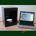
Automatic colony counting, inhibition zone determination, β - lactase detection, microscopic counting analysis system (HiCC IB combination type)
1. Product name: Fully automatic colony counting, inhibition zone determination, beta lactase detection, microscopic counting analysis system - HiCC-I
Product details
1. Name of goods:
Fully automatic colony counting, inhibition zone determination, β - lactase detection, microscopic counting analysis system - HiCC IB combination type
2 Main purposes:
Used for microbial colony counting analysis, inhibition zone measurement, antibiotic susceptibility potency analysis, sulbactam sensitive β - lactam enzyme assay, lactomycin potency analysis, and microscopic counting analysis, etc
3 System components:
Composed of a digital imaging system, analysis software, and computer.
4 Main Technical Performance Indicators
4.1 Imaging System
1)★Professional color digital 1/2.3 "photosensitive chip, true color 14 million pixels, resolution of 0.01mm, can distinguish colonies larger than 0.02mm/field of view 90mm diameter. Equipped with a 12mm non spherical optical lens;
2)★Microscopic imaging: Olympus CX31 (including a 10 megapixel camera), with a minimum resolution of 1um for microscopic analysis
3) Image capture: manual fine focusing, automatic/manual white balance, adjustable imaging brightness, automatic/manual color temperature control, parallel observation of dynamic and static images. Image enhancement, chromaticity adjustment, grayscale negative phase transformation, zooming in and out, field of view navigation.
4.2 Light source
1) Fully enclosed imaging environment;
2) The broadband LED lighting on the upper and lower sides easily achieves uniform backlight and floating dark field imaging effects.
4.3 Analyzed petri dishes, diameter
1) Analysis of culture dishes: pouring, coating, membrane filtration, spiral petri dishes, 3M paper sheets;
2) The diameter of the analyzed petri dish is 50-120mm (12mm non spherical optical lens).
4.4 Colony statistical characteristics
1) ★ Automatic recognition and statistics: SmartdownTM colony intelligent recognition technology automatically recognizes large-scale clustered and chain shaped adhered long rod-shaped bacteria (microscopic morphology of large adhered Escherichia coli, bacterial spores) and various types of colonies on petri dishes (including Staphylococcus aureus count (GB4789.10-2016), Escherichia coli group determination (GB4789.3-2016), etc.).Automatically generate batch processing guides to achieve one click automatic counting;
2) The automatic counting accuracy is ≥ 96.5%, with a maximum monitoring correction of 3.5%, achieving 100% accuracy. Analysis and statistics speed: 150-800 colonies/s;
3) ★ Colony adhesion segmentation: Automatically segment long, circular and other colony targets that adhere to each other in patches, and users can choose to segment or not segment. It is also possible to manually segment and merge colony target images;
4) ★ Color and shape recognition:Equipped with fast level set extraction technologyIt can identify and count the number of colonies of specific colors and shapes according to 20 categories, and can be corrected by category conversion. 6 target areas can be selected simultaneously for automatic analysis, and the analysis target areas can be freely increased, decreased, or edited;
5) Colony morphology analysis: Automatically obtain colony area, equivalent diameter, major axis, minor axis, aspect ratio, roundness, perimeter, shape factor, etc. Can be filtered and queried based on morphological indicators;
6) Click the mouse to correct and automatically remove impurities,Untrace removal of grids and textEffectively supports complex microbial statistics and complies with the parameter automatic conversion characteristics of GB4789.2-2016.
4.5 Measurement of Antibacterial Zone
1) One click fully automatic determination of all antibacterial zones in the petri dish, which can automatically determine the antibacterial zones with local adhesion. In the presence of local text interference, the antibacterial zone area, diameter, and other results can be automatically obtained. Measurement and analysis take 1-2 seconds, and can save or view various types of analysis data uniformly;
2)★The petri dish can be placed in any direction. It has the functions of automatic selection of antibacterial zone samples, accurate visualization comparison, and editing.The resolution error of the antibacterial zone measurement system is ≤ 0.001mm, and the repeated measurement error is ≤ 0.3%; The measurement error of potency is ≤ 0.3%; Repeated measurement error of potency ≤ 0.3%; Measurement difference between stations ≤ 0.2%;
3)★Compliant with the Chinese Pharmacopoeia, USP United States Pharmacopeia, and EP European Pharmacopoeia, capable of completing the latest National Pharmacopoeia (2020 edition) first, second, and third dose and combined calculations, as well as antibiotic potency analysis for all bacterial strains.Combination optimization analysis characteristics with potency;
4)★According to the food additive Streptococcus lactiae QB 2394-2007 standard, all inhibition zones on the petri dish can be automatically measured and the potency can be automatically analyzed;
5)★Built in American CLSI "Antimicrobial Susceptibility Test Execution Standard" latest 25th edition (2015)All data, providing a data input and modification platform for easy updates, providing a quick tool for paper based drug susceptibility analysis.
5)★The system administrator can assign usage permissions to more than 10 operators, and the operation records can be traced back to comply with GMP requirements.
4.6 Shubatan sensitive β - lactase test
1)★According to the national Ministry of Health's "Method for the Detection of Sulbactam Sensitive β - Lactamases in Milk and Dairy Products - Cup Plate Method", measure the content of sulbactam sensitive β - lactamases (Jinyulanase) in milk and dairy products.The diameter of the inhibition zone generated by tubes (A), (B), and (D) in the pure water control can be used to automatically determine whether the analysis results of the six petri dishes are valid. According to the diameter of the inhibition zone generated by tubes (A) and (B) in the sample, a positive or negative result output report can be automatically generated;
2) It has an automatic warning function for invalid reports, which can automatically remove impurities or defects based on differences in size, making the report more comprehensive and reliable. It can generate reports that comply with GLP/GMP requirements, adhere to the integrity of raw data and records, and provide comprehensive data support for laboratory certification. Historical data can be accessed freely, facilitating digital file management in the laboratory.
4.7 Microscopic Counting and Analysis Module
1) Image acquisition management: Capture images of active objects, save, open, edit, print, and browse them, and save them as static images in file formats such as Jpeg, BMP, PNG, TIFF, etc. It can continuously record the sequence of active target images or capture dynamic and static images at regular intervals, and store them in various formats such as AVI. All recorded images or image sequences can be easily played back, and any area of the active target within the microscope field of view can be dynamically zoomed in locally (with adjustable magnification);
2)★Real time preview saturation warningThe saturation warning feature during preview allows you to avoid capturing overexposed images;
3)★Real time puzzle and depth of field expansionAutomatically concatenate multiple views to obtain a complete image. Without the need for a Z-axis electric scanning table, images captured from multiple focal planes are automatically formed into multi focus depth extended 3D high-definition images, revealing image details;
4) Image observation and comparison: image scaling, rotation, mirroring, local observation, mesh mask, etc; Compare and contrast the processed image with the original image;
5) Selection of interest target area: Replace, merge, subtract, intersect any interest target area with circles, rectangles, polygons, and can be edited, deleted, or unselected;
6) Effect enhancement: Image brightness, contrast, saturation, RGB three color components, grayscale coefficient, erosion, dilation, hole filling, inverse adjustment, various histogram correction, smoothing, sharpening, denoising, edge detection, etc;
7) Color processing: automatic correction of light field non-uniformity (automatic background correction), color adjustment, image color separation and filtering, etc;
8) Image measurement: measuring line segments, multiple line segments, spline curves, arcs, circles, ellipses, rectangles, polygons, irregular shapes, lengths, areas, perimeters, angles, etc., with caliper tools. Online calibration of measurement units, magnetic lasso, vectorization of all shapes, editable, and arbitrary annotation on images (boundaries, numbers, symbols, fillers, etc.);
9)★Automatic segmentation of long or circular adhesive targets, automatic classification, counting, and measurement(Obtain morphological parameters such as area, perimeter, diameter, main axis, and secondary axis);
10)★Automatic learning classification feature based on color and shapeIt can interactively convert classification categories and has a classification enhancement function based on expert guidance for secondary self-learning;
11)★Sort output of target object(Provide corresponding distribution maps based on parameters such as area, size, aspect ratio, etc.), features also include: statistics by region or mouse click, querying or filtering, editing.
4.8 Measurement Function
1) Instrument calibration: automatic size calibration, also available for manual calibration
2) Automatic analysis: equivalent diameter, area, major and minor diameters, perimeter, roundness, shape factor, etc
3) Accurate measurement: can be dragged with the mouse to accurately measure length, angle, radian, area, arc, any curve, etc
4.9 Data Export, Report Printing, and Upgrading
1) The analysis of images and data results can be saved, and a total report will be automatically generated. The statistical analysis results will be output to an Excel spreadsheet
2) Online editing: can mark selected colonies
3)
Report printing: automatic printing of images and statistical data
4)
Software online automatic upgrade
5 Instrument specifications and configuration
1) 1 professional level color 14 megapixel camera and imaging host
2) Olympus CX31 three eye biological microscope (including 10 megapixel color microscope camera), 1 unit
3) Fully automatic colony counting analysis, antibacterial zone determination analysis, drug sensitivity potency analysis, β - lactase detection, microscopic counting analysis software and lock1 set4) Brand all-in-one computer (dual core CPU/4G memory/1G independent graphics card/hard drive 500GB)
Online inquiry
-
Contacts
-
Company
-
Telephone
-
Email
-
WeChat
-
Verification Code
-
Message Content
-