
VIP member

CKX53 Olympus Inverted Microscope CKX53
With improved image quality and ergonomics, the Olympus CKX53 provides performance and comfortable workflow requirements for various cell cultures, in
Product details
With improved image quality and ergonomics, the Olympus CKX53 provides performance and comfortable workflow requirements for various cell cultures, including live cell observation, cell sampling and processing, image capture, and fluorescence observation.
Observation of live cells
Olympus Inverted Microscope CKX53The high contrast achieved by the iPC system provides cells with a clear field of view at 4x, 10x, 20x, and 40x, without the need for users to switch or replace phase contrast rings. The new phase difference system facilitates faster and more comfortable workflow for simple and effective cell observation.

The PLN2X objective with a phase difference plate hole, CKX3-SLPAS, 22mm field of view, and an inner diameter of 11mm. The result is that the observation using this objective is perfectly effective in screening the desired cells, thereby achieving a faster cell culture process. The 2X objective provides significantly higher contrast, while other objectives enable clear identification of even transparent objects in the sample. For example, when viewing a 96 well microplate, a wide field of view allows for observation of all cells without the need to move the stage.

With this newly developed IVC technology, the phase contrast of the depth of field ratio enables the presentation of clear three-dimensional images for any shape or transparent object. In addition, the observation of IVC did not provide clear opinions on halo or directional shadows, and the completeness of the detailed information of the objects retained during the observation process.(Source: Chengguan Instrument)
*10X objective lens (PLCN10X, CACHN10XIPC) is used for the observation of this new IVC.


User oriented design for effective cell sampling and processing
The design of Olympus inverted microscope CKX53 is designed to fit on a clean mirror body. With its anti UV coating, the microscope can be left on a clean mirror body during the UV lamp sterilization process. Compared to previous models of CKX, the Olympus microscope CKX53 weighs about 7 kilograms and is lighter in weight, with a smaller volume that takes up less laboratory space. A microscope can be easily moved with just one hand and observed through the neck of the tube. The base of the microscope has a sliding pad for easy positioning.


The shorter distance between the viewpoint and the Olympus microscope CKX53 optical axis/focusing knob facilitates natural hand positioning, making focusing and cell sampling easier.

Whether observing from a standing or sitting position, the 45 degree angle of the eyepiece and the position of the butterfly shaped observation tube opposite the stage facilitate ergonomic cell observation. Aseptic work can begin and quickly complete the time outside the miniaturized cell culture box.(Source: Chengguan Instrument)
All including power switches, coarse and fine focal points, and knob controls for switching optical paths are ergonomically positioned to enhance operation and reduce user fatigue.

andOlympus Inverted Microscope CKX53The universal bracket is easily visible in various containers, including culture dishes, microplates, and flasks. When attaching the optional bracket, three additional 35mm culture dishes can be suitable for mounting on the stage. In addition, different types of microplates can be processed without retainers.
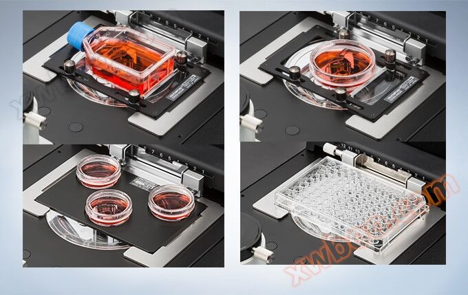
The width of the Olympus CKX53 microscope and the easily detachable spotlight can also view containers, such as multi-layer tissue flasks, up to a height of 190mm. The excellent depth of the focal point of the PLC4X objective enables quick and easy cell observation of the two bottom layers inside the multi-layer tissue flask.

The arm of the container handle allows users to manually position and lift the cell culture container. In addition, this stage can be extended to a greater processing flexibility of around 70 millimeters.
Fluorescence observation
The Olympus CKX53 inverted microscope standard fluorescence kit allows even weak fluorescence signals to be clearly observed with different integrated light sources, such as a 100W mercury lamp (U-LH100HG), a 130W high-pressure mercury lamp (U-HGLGPS), and third-party LEDs *. The same type of mirror device provided by our IX3 and BX3 microscopes can be set in the three slots of the reflector device slider.(Source: Chengguan Instrument)
*Not available in certain regions.
Designed for fluorescence observation using CKX53 on the "shading board". Shielding effectively blocks indoor light, improves the contrast of fluorescence, and enables clear fluorescence observation even under bright laboratory conditions. When using phase difference, the shading plate can lift the light transmitted to the sample.
CKX-CCSW software
CKX-CCSW software can automatically measure cell fusion and provide quantitative data recording, so researchers no longer need to rely on manual counting and fusion estimation. utilizeCKX53 microscopeThe software can count the number of cells and fusion percentage in the culture dish. This data can be saved as a CSV format file for easy export to a PC computer for evaluation and archiving.

Measurement of unstained cultured cells and fusion
Previously, the technique used to measure the number and density of cultured cells required staining the cells and manually counting them. Not all applications can perform specimen staining, such as regenerative medicine. Olympus CKX-CCSWsoftwareThe proprietary algorithm used enables the software to accurately count unstained cells in different containers. By regularly repeating this operation, users can create specific growth data for individual cells, which can be used to estimate the timing of the next cell channel when cell culture requires passage, in order to avoid overgrowth.

Reduce pollution risk
Living cells are typically cultured in a constant temperature incubator with appropriate temperature and CO2 concentration. During the cultivation process, it is sometimes necessary to remove cells from the incubator and check their growth status. By using microscope imaging, CKX-CCSW software can reliably determine the number of cells and culture density. This process is very rapid and can greatly shorten the time that the culture dish stays outside the constant temperature box, reducing the risk of contamination.
Quick and easy to operate
The interface of CKX-CCSW software is user-friendly, allowing users who have undergone brief training to quickly complete counting and fusion measurements. Specific calibration procedures have humanized characteristics, and measurement analysis based on multiple images can achieve higher counting accuracy.

Using quantitative analysis to improve the quality of cell culture process
Unlike manual cell counting and cell density estimation using a hemocytometer, CKX-CCSW software can provide reproducible quantitative growth data. This data can be saved as a CSV format file for easy archiving and recording. Both stacked image files displaying cell growth area and original image files can be saved in JPEG or TIFF format.


Specifications
project |
CKX53 |
||||
Model series |
bright field |
Difference Beginner |
Standard deviation |
fluorescence |
|
optical system |
UIS2 (Universal Infinite Distance Correction) Optical System |
||||
focusing |
Vertical motion system of coarse and fine focus knob objective converter for cyclic use. Travel: 20mm (Focus point: Top surface of flat stage up to 18.5mm) Per revolution stroke: 36.8mm (coarse), 0.2mm (fine) |
||||
Nosepiece |
manual |
Built in 4-hole position |
|||
stage |
Tablet carrier platform |
200mm (length) × 252mm (width) Merge and exchange transparent insertion boards |
|||
mechanical stage |
optional |
The microporous plate holder is equipped with anti detachment function at the XY coaxial knob on the right side of the loading platform on the tablet Platform travel: X=110mm, Y=74mm |
|||
Segmented loading platform |
70 mm(L) X 180 mm(W) |
||||
lighting system |
light source |
4000K color temperature LED light source |
|||
Filter holder |
Insert filters with a thickness of up to 6mm and ø 45mm, detachable |
||||
aperture diaphragm |
Aperture blades, manual opening/closing system |
||||
Insert slider |
optional |
Pocket with differential slider and built-in slider position, click stop mechanism(Source: Chengguan Instrument) Aperture of pre center iPC 4X, 10X, 20X, and 40X The insertion direction can be adjusted within a range of ± 30 degrees to the right or left |
|||
IPC slider |
optional |
Pre center difference aperture 4X, 10X, 20X, 40X and 2 ø 45mm holes |
|||
condenser |
Large numerical aperture NA: 0.3 Working distance WD: 72mm Applicable objective magnification of 2X, 4X, 10X, 20X, and 40X The tissue bottle with a height of up to 190 millimeters can be loaded with a non removable spotlight on the stage(Source: Chengguan Instrument) |
||||
observing tube |
Fixed binocular tube with a tilt angle of 45 degrees Pupil distance 48-75mm Optical path: eyepiece/camera port=100/0 0/100 |
||||
Camera port |
Olympus camera adapter interface |
||||
eyepiece |
Magnification: 10X FN22 |
||||
fluorescent |
optional |
Detachable lighting 3CH switching slider |
|||
FL light source |
100W mercury lamp |
||||
FL optical shutter |
available |
||||
FL Field of View Station |
available |
||||
FL excitation block |
2 excitation blocks (B&G) and UIS2 mirror device (optional) |
||||
FL shading board |
A sunshade can prevent light from indoors |
||||
Overall dimensions |
200 (W) x 498 (D) x 454 (H) mm (different configuration) |
||||
weight |
About 6.9 kg(Source: Chengguan Instrument) |
||||
Rating |
AC 100-240V 50/60 Hz 0.4A |
||||
power consumption |
Less than 4W |
||||
operating environment |
ambient temperature |
Indoor temperature range of 5-40 ℃ (41-104 ℉) |
|||
High relative humidity |
When 80% of the indoor temperature reaches 31 ℃ (88 ℉), 70% reaches 34 ℃ (93 ℉), 60% reaches 37 ℃ (99 ℉), and 50% reaches 40 ℃ (104 ℉)
|
||||
Outline dimension image:

Online inquiry
-
Contacts
-
Company
-
Telephone
-
Email
-
WeChat
-
Verification Code
-
Message Content
-